
Blog | bellevue Podiatry

Toe Walking Podiatrist Rosanna: Will Your Child Grow Out of It?

Toe Walking Podiatrist Rosanna: Will Your Child Grow Out of It?

Many parents notice their child walking on tiptoe, heels barely touching the ground, and wonder whether it’ll sort itself out. In most cases it does: children who toe walk on both feet with no medical cause, known as idiopathic toe walking, usually settle into a normal heel-to-toe gait by about age 10.

What matters more than age is whether your child can still get their heels flat, and whether there’s pain, toe walking on one side only, tight calf muscles or Achilles tendons, or frequent tripping or falls. Those signs tell you whether simply watching is fine or a proper assessment is the better next step.

In This Article

  • Will they grow out of it: What a Swedish study found about how many children stop toe walking on their own by age 10.
  • Signs worth getting checked: The red flags, like heels that won’t sit flat or toe walking on one side, that mean it’s time for an assessment.
  • What the podiatrist checks: How we test ankle movement, calf tightness and walking pattern, and what happens once the assessment is done.
  • Treatment and the evidence: From home stretches and gait plates to casting, and why treatment is matched to your child’s actual problem, not their age.
  • Help close to home: How families in Rosanna and Melbourne’s north-east can get toe walking checked, and what the appointment involves.

Will My Child Grow Out of Toe Walking?

Will they grow out of it?
Usually yes, as long as the heels can still get down
4 in 5
children who had ever toe walked had stopped on their own by age 10, with no ankle stiffness (Swedish study, 1,401 children).
1 in 20
healthy children toe walk at some stage. It’s common, so seeing it on its own isn’t a cause for alarm.
No pain + no stiffness
means no treatment is needed, according to a 2024 UK consensus of children’s orthopaedic surgeons and physiotherapists.
The check that matters
Can your child stand with heels flat and walk on their heels? If not, or if it’s one side only, have it assessed rather than waiting.
The odds of a child growing out of idiopathic toe walking, and the heel-flat check that tells you whether waiting is still reasonable.

Hearing “they’ll grow out of it” for a few years can start to feel like a brush-off, but for most children it’s backed by research. A Swedish study of 1,401 children found around 5% of healthy kids toe walk at some stage, and by age 10 four in five had stopped on their own, without developing ankle stiffness.

That outlook applies to idiopathic toe walking, meaning toe walking on both feet with no underlying medical cause, which often runs in families, so you may spot it in a parent or sibling too.

The same study found a small group whose ankles were already stiff early on, and the researchers treat that as a separate problem, a short Achilles tendon, that’s better managed early in childhood. So the useful question isn’t how old your child is, but whether they can still get their heels flat.

A 2024 UK consensus of children’s orthopaedic surgeons and physiotherapists agreed that toe walking without pain or ankle stiffness doesn’t need treatment. You can do a quick version of the heel check at home (see the FAQ below), and it’s the first thing we test in clinic.

When Is Toe Walking Worth Getting Checked?

Young child standing barefoot with heels lifted off the floor during a toe walking check

Seek professional advice if your child cannot comfortably stand with their heels resting flat on the ground.

For many Victorian families, the first prompt comes from the Maternal and Child Health 3.5-year check, a kinder teacher or your GP. In my experience, it’s wise to follow this up calmly rather than dismiss it, because an early assessment gives you a clear baseline to compare against later.

The Royal Children’s Hospital Melbourne lists these red flags as reasons for further referral:

& Can’t stand with heels flat on the ground or walk on their heels

& Toe walking on one side only

& Enlarged calf muscles

& Frequent tripping and falling

& Signs of an underlying nerve or muscle condition

A toe walking assessment is also worthwhile if there’s foot or leg pain or the way your child walks is affecting their confidence or play, and it doesn’t automatically mean treatment.

Toe walking that hangs on is more common in children with developmental differences such as autism or ADHD, and the Swedish researchers found these were common among children still toe walking at 10. Because one in 20 otherwise healthy children toe walk too, it isn’t a reason to assume anything more on its own, so mention any other developmental concerns when you book and we’ll look at the whole picture.

What Does a Toe Walking Podiatrist Check?

Podiatrist reviewing a tablet video of a child's walking pattern during a toe walking assessment

Video gait analysis allows us to slow down and review exactly how your child's feet contact the ground during movement.

When families see a toe walking podiatrist in Rosanna, we start with a thorough history: your child’s developmental milestones, whether toe walking runs in the family, and anything kinder or school staff have noticed.

From there, a biomechanical assessment for children typically looks at four things:

  • Ankle range of motion: We check whether your child’s heels can rest flat and how far the ankle bends upwards (dorsiflexion), with the knee both straight and bent.
  • Calf muscles: One calf muscle (gastrocnemius) crosses the knee and the other (soleus) doesn’t, so comparing those two positions shows which one is tight. We also check strength and whether the calves look enlarged.
  • Walking pattern: We complete a children’s gait assessment in our Rosanna clinic, often using video, so you can see exactly how each foot meets the ground.
  • Balance and neurological signs: We note frequent trips or falls, one-sided toe walking or changes in reflexes, as these may point to an underlying cause.

Once the assessment’s done, we’ll explain what we found and whether monitoring, treatment or a referral to another health professional makes most sense. If anything suggests a nerve, muscle or developmental cause, we’ll say so plainly and suggest you see your GP rather than guess.

Toe Walking Treatment and What the Evidence Says

Matching treatment to the problem
How toe walking treatment usually steps up
No single treatment has strong trial evidence, so each step has a goal and a point where we review it.
1
Assess
Heel-flat and heel-walking check, ankle bend with the knee straight and bent, gait and a basic neurological screen.
2
Ankle moves freely
No pain or stiffness usually means no treatment is needed. Home stretches, heel-walking games and supportive shoes or gait plates are optional extras.
3
Ankle won’t reach flat
Serial casting to regain range, then night splints to hold it, with an agreed goal such as heels flat by the end of casting.
4
Goal not met
Referral through your GP to a paediatric orthopaedic surgeon or paediatrician to talk through the next options.
A four-step flow showing how toe walking treatment is matched to whether the ankle moves freely, with a goal and review point at each step.

If treatment is suggested, it’ll usually aim to ease pain or ankle stiffness, or to support your child’s confidence and play, rather than simply change how they walk.

Options range from calf stretches and strengthening at home, to supportive shoes and stiff full-length insoles called gait plates, through to casting, night splints or a referral to a paediatrician or orthopaedic surgeon. Which one fits depends on whether the problem is mainly habit or a genuinely stiff ankle, and that’s what the assessment tells us.

Honest answers matter here, because a 2019 Cochrane review found just four small trials (104 children in total) and said the evidence was too weak to draw conclusions about any treatment. So rather than open-ended treatment, we agree a clear goal and a timeframe with you, and change course if it isn’t being met.

Calf Stretches and Strengthening at Home

When the calf muscles and Achilles tendon are tight, it’s physically harder for a child to bring their heels to the ground. That’s why gentle stretching and strengthening at home is often the first step, and the Royal Children’s Hospital fact sheet notes that daily calf stretches may help.

Simple toe walking exercises families can start with include:

  • Wall stretch: Hands on the wall, one foot stepped back with the heel flat, held for 30 seconds. Repeat with the back knee bent to reach the deeper calf muscle.
  • Heel walking: A “penguin walk” race down the hallway works the muscles at the front of the shin.
  • Flat-heeled squats: Have your child squat with their heels down while playing with toys on the floor.
  • Step stretch: With your child holding the rail, let their heels lower gently off the bottom stair.
  • Balance games: Standing on one leg, or walking heel-to-toe along a line, forwards or backwards.

Keep sessions short, playful and part of the daily routine, stop if your child reports pain, and pair them with the right footwear or orthotics for toe walking where needed.

Shoes, Insoles and Gait Plates

Alongside stretches, the shoes your child wears every day can quietly influence their walking pattern. Supportive shoes for children who toe walk typically have:

  • A firm heel counter to hold the heel securely in place
  • A supportive sole that resists excessive bending
  • A secure fastening, such as laces or Velcro

Thongs and soft, flexible shoes aren’t thought to cause toe walking, but they give little resistance to a child who likes being up on their toes. Gait plates, stiff full-length insoles worn inside the shoe, are used to encourage a heel-first step, possibly through extra stability and feedback along the whole sole.

In a small Australian trial of 15 children, rigid full-length insoles worn inside high-top boots increased heel contact straight away. Whether that lasts long term isn’t yet known, so we treat footwear as one part of a plan rather than a fix on its own.

Casting, Night Splints and Referral Options

If stretching and footwear haven’t improved your child’s toe walking, or their ankles have become stiff, we may discuss options that aim to restore ankle dorsiflexion, the ankle’s ability to bend the foot up towards the shin.

  • Serial casting: A series of below-knee casts, changed over several weeks, gradually lengthens tight calf muscles and the Achilles tendon.
  • Night splints: A brace worn during sleep that holds the ankle in a gentle stretch to help keep the range gained.
  • Paediatrician referral: We may recommend this to rule out neurological, muscular or developmental causes of toe walking.
  • Orthopaedic surgeon referral: Generally considered when ankle stiffness persists despite non-surgical treatment.

The 2024 UK consensus suggests agreeing a goal up front, such as heels flat after about six weeks of casting, and seeking a surgical opinion if that goal isn’t reached. With so little trial evidence behind any option, as the Cochrane review found, that goal-and-review approach is what stops months of treatment that isn’t working.

Whether you’re looking for a toe walking podiatrist near Greensborough or help closer to Preston, we’ll explain each option clearly and decide the next step together.

Toe Walking Help in Rosanna and Melbourne’s North-East
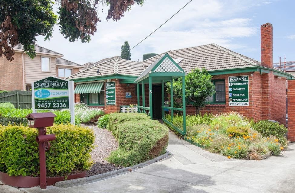
Modern suburban clinic building on a leafy street in Rosanna, Melbourne

Local assessments provide clear answers for families in Rosanna and surrounding Melbourne suburbs without needing distant travel.

If your Maternal and Child Health nurse, kinder teacher or school has raised your child’s toe walking, a local assessment can give you a clear answer without travelling far.

Bellevue Podiatry, at 8 Bellevue Avenue, Rosanna, sees children from Heidelberg, Ivanhoe, Macleod, Viewbank, Watsonia, Preston, Bulleen, Greensborough, Montmorency and Lower Plenty.

Many young children walk on their toes while they’re learning, and that’s a normal part of development. Most have settled into a heel-to-toe pattern by about age two, so toe walking well past that, or alongside other concerns, is worth having looked at.

The appointment is relaxed and hands-on: we watch your child walk and run, gently check their feet, calves and ankles, and look at how toe walking affects their balance, play and everyday activities. Many children treat it like a game, which helps us see how they really move.

Whether you’ve noticed a toddler walking on their toes or an older child who’s kept the habit, you’ll leave knowing what’s going on and which options, if any, make sense.

We’ll also tell you if a referral to your GP or a paediatrician is worth considering.

Frequently Asked Questions

Should I Keep Reminding My Child to Put Their Heels Down?

In most cases, constant reminders aren’t needed. In practice, frequent prompting rarely shifts a toe walking habit on its own and can leave your child feeling self-conscious, which may dent their confidence and enjoyment of play.

An occasional, gentle reminder is perfectly fine.

What matters more is whether your child is physically able to bring their heels down when asked.

What Can I Check at Home?

Ask your child to stand with both heels flat on the floor and their body upright, then walk a few steps on their heels. These are the same quick checks the Royal Children’s Hospital suggests GPs use before deciding on referral.

If they can do both comfortably, it usually means there’s enough flexibility in the calf muscles and Achilles tendons for now.

It’s worth having their feet, calves and gait (walking pattern) assessed if they can’t do either, if they toe walk on one side only, or if they trip or fall a lot.

An assessment helps us identify any muscle tightness, joint restriction or other underlying cause early. From there, we can suggest management that suits your child.

Did I Cause My Child’s Toe Walking by Using a Baby Walker?

Probably not: there’s little research on baby walkers and toe walking, but idiopathic toe walking commonly runs in families, which points away from anything you did.

“Idiopathic” simply means no medical cause can be found, so this isn’t something to feel responsible for. The more useful thing to check now is whether their heels can rest flat, since that tells you more than how it started.

Do I Need a GP Referral to See a Podiatrist About Toe Walking?

No, you don’t need a GP referral to see a podiatrist about toe walking. You can book directly online or call us on (03) 9457 2336.

Many families come to us after a kinder teacher, Maternal and Child Health nurse or GP raises it, but noticing it yourself is reason enough to have it checked.

If anything needs a closer look, we’ll let you know, and your GP can refer you to a paediatrician for further investigation.

Does Going Barefoot or Wearing Thongs Make Toe Walking Worse?

There’s no strong evidence that going barefoot or wearing thongs causes toe walking or makes it worse. Parents often worry about this, but footwear alone isn’t considered a main cause.

For most children, barefoot play is fine, and walking on varied surfaces lets the feet move naturally and build strength.

Why Podiatrists Still Talk About Footwear

Even though footwear isn’t thought to cause toe walking, the right shoes can be part of a management plan. Supportive shoes, and sometimes stiff full-length insoles called gait plates, may help your child settle into a heel-first walking pattern.

They’re tailored to your child and usually used alongside other strategies rather than as a standalone fix.

When to Have Your Child Assessed

If you’re unsure whether your child’s walking needs attention, a podiatry assessment can give you clarity. At Bellevue Podiatry, we look at your child’s feet, calf muscles and gait to understand what’s behind their toe walking.

This is general information only and doesn’t replace an individual assessment of your child.

How Can I Prepare My Child for Their First Podiatry Appointment?

Keep it simple and reassuring: tell your child they’ll meet a friendly podiatrist who’ll watch them walk and check their feet and legs. Calling it a relaxed check-up helps ease nerves and lets us see how they naturally move.

To help with the gait assessment, please bring:

  • Shorts for your child to wear, so we can clearly see their knees, calves and ankles
  • The shoes they wear most days, as wear patterns offer useful clinical clues
  • Notes on when you first noticed the toe walking and whether it runs in the family
  • Any observations from kinder, school or your Maternal and Child Health nurse
  • A short home video of your child walking barefoot

The video is especially useful because children often walk differently in an unfamiliar clinic. Seeing their usual movement at home gives us a truer picture of their walking pattern.

Conclusion

If your child’s toe walking has you worried, take heart: about four in five idiopathic toe walkers in the Swedish study had stopped by age 10. Not every child does, though, and heels that won’t sit flat, one-sided toe walking, tight calves or frequent tripping are better assessed than met with a wait-and-see approach.

At Bellevue Podiatry, we help families from Rosanna, Heidelberg, Watsonia, Ivanhoe and nearby suburbs work out where their child stands, with an evidence-based approach that’s honest about what research can and can’t tell us. Book an assessment online or call (03) 9457 2336, and we’ll help you decide whether to watch, treat or refer.

Your child still walks on their toes, and you've been told they'll grow out of it? Most do — but the real question is whether their heels can still sit flat.
Call our friendly Reception on (03) 9457 2336 or book online for a toe walking assessment in Rosanna — we check your child's ankle range and how they walk and run, so you know whether to keep watching or take the next step.
Mention code NEW85 — initial podiatry consultation $85 (normally $110), new patients only.

Will They Grow Out of It?

A one-minute heel check you can do at home — plus what the research says, the signs worth acting on, and what treatment actually involves.

Foot Stress Fracture: Warning Signs, The Right Scan And Real Recovery Times

Foot Stress Fracture: Warning Signs, The Right Scan And Real Recovery Times

A stress fracture in the foot rarely comes from one bad step. It builds up gradually from repetitive loading – training sessions, long walks, or high-impact work that outpaces the bone’s ability to repair itself.

Here’s the short answer. A foot stress fracture usually shows up as a dull, localised ache at one exact point on the bone that worsens the longer you’re on it, and an early X-ray is often clear even when the injury is real – so the diagnosis rests on how the pain behaves, backed by an MRI when the picture still doesn’t add up.

Plenty of people put it down to worn-out footwear or a harder training surface, when what’s going on is a genuine bone injury. If foot pain has hung around more than a few days without shifting, have it looked at properly.

In This Article

  • How the injury builds: why training load outruns your bone’s repair, and what a stress reaction is before it becomes a fracture.
  • The checks you can do tonight: pinpoint bone tenderness, the hop test, and the signs that mean you shouldn’t sit on it.
  • Why a clear X-ray isn’t the all-clear: what plain film misses early, what an MRI adds, and what Medicare actually covers here.
  • Why the exact bone matters: the foot bones that need cautious handling, and the ones that usually settle with load management.
  • Offloading and getting back to sport: what the moon boot is actually for, and the milestones that decide when it comes off.

How A Foot Stress Fracture Actually Starts

How bone stress builds up

Each stage is the same injury, further along. Nothing snaps at the start.

Stage 1

Load climbs

More kilometres, more court time, harder surfaces, less recovery between sessions.

→

Stage 2

Repair falls behind

Old bone is cleared away faster than new bone can be laid down.

→

Stage 3

Stress reaction

The bone is irritated and sore under load, with no visible crack yet.

→

Stage 4

Stress fracture

The bone gives way at its weakest point and pain starts limiting you.

The catch: plain X-rays generally can’t show stages 1 to 3, and often not the first couple of weeks of stage 4. A clear film early on doesn’t rule any of this out.

Four-stage flow showing how repeated load outruns bone repair, and why early X-rays commonly look normal.

A stress fracture doesn’t switch on overnight – it builds up quietly, session by session.

Every time you run or land from a jump, your foot bones flex slightly and rebuild themselves in the background. That rebuilding is a race, and repeated loading speeds up the clearing side of it, so old bone comes away faster than new bone goes down.

When training load climbs faster than that repair can keep up – more kilometres, more court time, less recovery – the bone starts losing the race. That’s a stress reaction, the early and quieter stage of a bone stress injury.

Left unmanaged, a stress reaction can progress to a genuine foot stress fracture.

Which is why an ache that “comes and goes” after netball or a long run deserves attention early, rather than once it’s stopped you training altogether. The sooner it’s caught, the lower down that flow you tend to be.

What A Foot Stress Fracture Feels Like, And The Checks You Can Do Tonight

Podiatrist pressing one spot on a patient's midfoot to check for pinpoint bone tenderness

Pinpoint bone tenderness that worsens with activity helps separate bone stress from a soft tissue injury.

The pain pattern is the most useful information you have before any scan.

Bone tends to hurt at one exact point and stay sharp under load, while a tight calf or an irritated tendon usually spreads across a wider area and eases off once you’re warm.

Below are the checks worth running tonight, plus the handful of signs that mean skipping the wait-and-see approach and getting seen.

The Pain Pattern That Points To Bone Rather Than Soft Tissue

In clinic we often ask people to press along the sore area with one finger. If you land on pinpoint bone tenderness – a small, exact spot rather than a general ache – that’s worth taking seriously.

Soft tissue problems, like a strained tendon, tend to hurt across a broader area and typically settle once you’ve warmed up.

Bone pain often behaves differently, staying sharp and localised even once you’re moving.

The hop test adds another piece. Sharp, focal pain when you hop on the sore foot suggests the bone is struggling under load, though it’s worth knowing the test hasn’t been well studied – so treat it as a clue, not a verdict, and the same goes for a foot stress fracture x-ray taken early.

Three Simple Self-Checks Before You Book Anything

These three checks won’t give you a diagnosis. What they will do is help you describe the problem accurately when you come in, which genuinely speeds things up.

  • One-leg hop test. Stand on the sore foot and try a small hop. Sharp, localised pain is one of the classic foot stress fracture symptoms.
  • Point with one finger. Press firmly along the bone. A metatarsal stress fracture often hurts at one exact spot, not across a general area.
  • Rest response. Note whether the pain settles within minutes of stopping, or lingers for hours afterwards.

Write down what you find. It’s useful clinical information, and it feeds into whether an MRI for foot stress fracture is the sensible next step.

Signs That Mean You Should Be Seen Straight Away

Some presentations shouldn’t wait for a booking that suits your calendar. A few red flags change the timeline, so book in soon if you notice any of the following:

& You’re limping or altering your gait to avoid pain

& Pain wakes you at night

& Pressing on one specific spot on the top of your foot reproduces the pain sharply

Separately, a few things move this out of podiatry territory: you can’t put weight through the foot at all, the foot looks misshapen, or you’ve lost feeling in it. Any of those needs same-day medical assessment.

Pain over the midfoot can point to a navicular stress fracture, one of the higher-risk injuries we treat cautiously because it doesn’t always show up well on a standard X-ray. In that situation an MRI is usually the more informative next step, and a bone scan is an alternative where MRI isn’t available.

In the meantime, a cam walker boot can take load off the area and keep you moving while the diagnosis is sorted out.

Why Your X-Ray Can Come Back Clear And You Still Have A Stress Fracture

Podiatrist comparing a clear foot X-ray with MRI images showing early bone stress changes

Early stress fractures often don’t show on X-rays, while an MRI can pick up bone stress changes sooner.

A clear X-ray does not rule out a stress fracture.

Bone stress sits on a continuum, and in the early stages the plain film simply has nothing to show yet.

& Timing is the main reason. X-rays only reveal change once the bone starts laying down new healing tissue, which takes two to three weeks to become evident, whereas MRI is reported as far more sensitive to early bone stress.

& Location also plays a part. A fifth metatarsal stress fracture or a navicular injury can stay subtle on imaging while your symptoms are perfectly clear, and it’s well recognised that these cracks are hard to see on an X-ray at first.

& Better imaging exists, but access isn’t even. An MRI picks up bone marrow changes far earlier than an X-ray, making it the more reliable tool when a stress fracture is suspected and unconfirmed.

Here’s where the Australian system shapes the plan rather than the textbook. Under Medicare, the imaging a podiatrist can request is limited to a specific list of foot, ankle and leg X-ray and ultrasound items – MRI and CT need a medical practitioner’s request.

Put those two facts side by side and the practical conclusion is uncomfortable but useful. The test we can arrange fastest and most cheaply is the one least likely to find an early injury, so the first few weeks of management are built on how your foot behaves, not on what a film shows.

That’s why a normal X-ray at week two shouldn’t change your plan.

If the clinical picture says bone, we treat it as bone and review it.

In practice around Melbourne’s north-east that usually looks like this: we assess, request a foot X-ray or ultrasound ourselves where it’s genuinely useful, start offloading straight away, and work with your GP if an MRI is warranted. Worth asking the imaging practice for a quote first, because foot and ankle MRI often carries an out-of-pocket cost.

Not All Foot Stress Fractures Are Equal: High-Risk Versus Low-Risk Bones

Not every foot bone behaves the same way

Same injury, different bone, different plan. This is the first thing we want to pin down.

Usually lower risk

Tend to settle with load management

Second, third and fourth metatarsal shafts
The heel bone (calcaneus)
The fibula, at the outside of the ankle

Decent blood supply, load spread across neighbouring bones.

Higher risk

Handled more cautiously, reviewed more closely

The navicular, in the midfoot
The base of the fifth metatarsal
The talus
The sesamoids, under the big toe
The first metatarsal

Poorer blood supply or high, concentrated load – slower to settle.

Two dials, not one: which bone, and how far the injury has progressed. In pooled research across bone stress injuries, average time back to sport ran from roughly six weeks for the mildest grades to around fourteen weeks for the most advanced – which is why “six to eight weeks” is a starting point rather than an answer.

Comparison panel of lower-risk and higher-risk foot bones, with the two factors that set a recovery timeline.

Once we’ve established there’s bone stress in your foot, the next question is where it sits. That answer changes how carefully we manage it.

Foot stress fractures are grouped as higher or lower risk depending on the bone involved, and the grouping drives the plan.

The split comes down to blood supply and how concentrated the load is. A metatarsal shaft shares its work with the bones either side of it and tends to settle predictably, while the navicular or the base of the fifth metatarsal does neither – so it gets offloaded for longer and reviewed more closely.

A sesamoid stress fracture under the big toe joint is a good example, because it needs more cautious handling than a metatarsal shaft – it’s carrying load every time you push off.

Grade matters as much as location. Pooled return-to-sport times rise steadily with the severity seen on MRI, averaging around 42 days at the mildest grade and around 99 days at the most advanced.

Location and severity move your timeline independently of each other. A higher-risk bone caught early can be a shorter road back than a lower-risk bone left to run for a season.

Which is why identifying the exact bone, and how far along it is, matters as much as identifying the injury itself.

Offloading, The Moon Boot, And Getting Back To Running Or Netball

Podiatrist fitting a grey CAM walker moon boot to an athletic patient's foot in clinic

A CAM walker can take load off a higher-risk bone while it settles, without putting your whole week on hold.

Once we know which bone is involved and how irritable it is, the next decision is how much weight it can safely carry. That decision shapes your foot stress fracture treatment plan more than anything else.

A boot isn’t rest. It’s a way of keeping you moving while one specific bone carries less – which is why it should come with an exit plan from the day it goes on.

Higher-risk bones and more painful presentations often call for a moon boot (CAM walker). It isn’t just a comfort measure either – when researchers measured pressure at the base of the fifth metatarsal, the CAM boot produced measurably less pressure than a post-op sandal or a normal shoe.

That’s the useful part. It offloads the sore bone while you keep walking through your day, so you’re not trading a bone injury for six weeks of lost fitness. It also means time in the boot is a decision, not a default. We aim for the least you need to get to comfortable walking, then we start taking it off.

Getting back to running, or back onto the netball court, follows a graded plan rather than a fixed date. There are three stages we typically work through:

  • Pain-free daily activity out of the boot
  • Walking, then jogging, building time and distance gradually
  • Sport-specific drills, then full training

Each stage has to hold up before the next one starts. Alongside it we review your footwear, orthotic support and training load, because the load that caused this is still waiting for you at the end.

Plenty of the bone stress we see comes off the same few surfaces – hard netball courts in winter, a pre-season block crammed into three weeks, and the Main Yarra Trail. A fair number of those people reach us through the clubs around Heidelberg and Ivanhoe.

Frequently Asked Questions

Does Medicare Cover the X-Ray or Ultrasound Referral for This?

For X-ray and ultrasound of the foot, ankle and leg, generally yes. Podiatrists can request a defined list of these items with a Medicare benefit attached, so we can often arrange that part ourselves on the day.

MRI and CT work differently. Those requests have to come from a medical practitioner, and foot and ankle MRI frequently carries an out-of-pocket cost, so it’s worth asking the imaging practice what you’ll pay before you book.

We can talk you through the options at your assessment, or give us a call on (03) 9457 2336.

Can I Still Do Gym or Swimming While My Bone Heals?

Often, yes – provided the activity doesn’t load the affected bone. That’s the whole test.

Swimming and pool-based exercise usually stay comfortable, and upper-body or seated gym work is typically fine too.

Impact machines, running and standing weights may need to wait until we’ve confirmed things are settling.

We’ll tailor this to your imaging and the specific location of the bone stress injury. Book online or call (03) 9457 2336 and we’ll map out what you can keep doing.

Does My Diet or Fuelling Affect Stress Fracture Risk?

Yes, and it’s the part most people skip past. If you’re training for a spring running series while skipping meals to fit the sessions in, low overall energy intake along with calcium and vitamin D shortfalls can leave bone less able to cope with repeated load.

Under-fuelling matters more than most athletes expect. International consensus on relative energy deficiency in sport describes impaired bone health as one of the most consistently documented consequences of not eating enough for your training load.

None of this is about eating perfectly. It’s worth raising with us, your GP or a dietitian, particularly if:

& Periods have become irregular or stopped

& Energy levels feel persistently flat

& Training load has climbed without any matching increase in food

Will Custom Orthotics Stop This Happening Again Next Season?

Orthotics can help. They’re not insurance against another injury, though, and it’s worth being straight about that.

They work by changing how load travels through your foot, which may ease pressure on the bone that was sore. They help most alongside decent footwear, sensible training progression and adequate fuelling, not instead of them.

We can assess your foot posture and gait to see whether orthotics genuinely suit your pattern before you commit to a pair.

How Soon Should I Book an Assessment After Symptoms Start?

If a niggle in your foot or shin hangs around beyond three to four days without settling, book it in rather than wait and hope.

Early review lets us arrange the right imaging and start offloading the affected bone sooner. Since time back to sport tends to track the severity of the injury on imaging, being further down that scale when you’re first seen is generally in your favour.

Leaving it can allow a low-grade stress reaction to progress, and higher grades typically mean a longer road back.

Call Bellevue Podiatry on (03) 9457 2336 or book online. Quote NEW85 for an $85 initial consultation, normally $110 – new patients only, one per person, valid 14 days.

Conclusion

A sore foot that keeps getting worse every week is telling you something, and rest alone rarely settles the argument. If you can press one spot and reproduce the pain sharply, or you’re still limping after a few quiet days, that’s the point to get it assessed rather than guess.

Don’t let a clear X-ray talk you out of it either. Respect the boot, follow a graded return, and let the milestones set the pace instead of the calendar.

At Bellevue Podiatry we work from current clinical evidence and build each recovery plan around your assessment, your sport and the bone that’s actually involved. We see people from Rosanna, Heidelberg, Watsonia, Ivanhoe and the surrounding suburbs, and we’d rather look at a niggle early than a fracture late.

Book an assessment online or call (03) 9457 2336. Quote NEW85 for an $85 initial consultation, normally $110 – new patients only, one per person, valid 14 days.

Sore foot getting worse every week, and the X-ray came back clear? An early X-ray often misses a stress fracture.
Call our friendly Reception on (03) 9457 2336 or book online for a foot stress fracture assessment in Rosanna — we check the bone directly, so you know where you stand before your next run or game.
Mention code NEW85 — initial assessment $85 (normally $110), new patients only.

Is My Sore Foot a Stress Fracture?

A three-minute check you can do at home — plus why a clear X-ray does not rule it out, and what getting back to running or netball actually looks like.

Plantar Plate Tear: The Forefoot Injury Often Mistaken for a Neuroma

Plantar Plate Tear: The Forefoot Injury Often Mistaken for a Neuroma

Plenty of people arrive at Bellevue Podiatry having already been told they have a neuroma, only to find the treatment hasn’t touched the pain. The burning or tingling they were warned about never quite turns up.

Instead there’s a stone-like ache directly under the joint, and sometimes a toe that has started to sit slightly out of line.

That pattern often points somewhere else entirely: a plantar plate tear. Before you persist with metatarsal pads or neuroma-specific treatment that isn’t helping, it’s worth having the joint itself assessed, because treating the wrong structure delays the right care and gives the real problem time to progress.

In This Article

  • Why the two get confused: Plantar plate tears and Morton’s neuroma sit in the same patch of forefoot, which is why one gets mistaken for the other.
  • What the plantar plate actually does: How a small ligament under your toe joint holds the toe down, and what changes when it stretches or tears.
  • Why pads and rest have not worked: The reason general forefoot remedies leave a torn ligament and a drifting toe exactly where they found them.
  • How the diagnosis gets confirmed: The hands-on tests and imaging used to separate a plantar plate tear from a nerve problem.
  • What treatment involves: How offloading, taping and custom orthotics take pressure off the joint so it can settle.

Why a Plantar Plate Tear Is So Often Mistaken for a Neuroma

Same spot, different structure: how the two conditions differ

Plantar plate tear

Structure: ligament under the toe joint

Pain feels like: a stone under the joint that never moves

Usual location: under the second toe joint

Worse with: push-off, barefoot walking

Visible sign: toe drifting sideways or sitting raised

Morton’s neuroma

Structure: thickened nerve between the toe bones

Pain feels like: burning or electric shock

Usual location: between the third and fourth toes

Worse with: tight or narrow shoes

Visible sign: usually none, toes look normal

The overlap: a torn plantar plate can irritate the nearby nerve and produce neuroma-like burning, which is how the wrong diagnosis gets made.

Comparison of plantar plate tear and Morton’s neuroma by structure, pain quality, location and visible signs.

Both problems live in the same small patch at the ball of your foot, so the overlap in symptoms makes sense. A 2025 narrative review of plantar plate diagnosis describes the injury as frequently underdiagnosed, with a presentation that overlaps Morton’s neuroma and joint inflammation.

A neuroma is a thickened nerve sitting between the toe bones. A plantar plate tear involves the small ligament under the toe joint that holds the toe down and pointing forward.

Because the ache, the swelling and the forefoot pain feel so alike, it helps to know what tends to separate them.

What a Plantar Plate Tear Tends to Feel Like

A plantar plate tear turns up in the same place as a neuroma, the ball of the foot, just beneath the toes. The structure involved and the pattern of symptoms, though, are different.

Pain that worsens as you push off is the usual complaint, often described as standing on a small stone that never moves. It tends to be worse barefoot and easier in a firm shoe.

The second toe is the one most often affected, and you may notice it drifting sideways or sitting slightly raised, as though it has lifted away from the ground.

Some people also see mild swelling on top of the foot. Others describe an odd sensation of a sock bunched up under the toes when there’s nothing there at all.

What a Morton’s Neuroma Tends to Feel Like

In clinic, Morton’s neuroma tends to be described quite differently:

  • Burning or electric-shock pain, most often in the third web space between the third and fourth toes
  • Numbness or pins and needles that come and go
  • A sense of something rolling under the foot
  • Relief from taking the shoe off and rubbing the area

Here’s the catch. A plantar plate tear can produce a pseudoneuroma sign on imaging and mimic those same nerve symptoms.

That overlap is precisely why a misdiagnosed neuroma is so common when the real problem is structural.

Simple Checks You Can Do at Home Before Your Appointment

A few simple checks at home can tell you whether your symptoms lean towards a plantar plate tear rather than a neuroma. None of them settle the diagnosis, but they’re worth doing before you book.

  • Visual check: Stand barefoot and look down at your toes. If one sits slightly raised or drifts sideways, that’s worth noting.
  • Palpation check: Gently press under the ball of your foot, just behind the second and third toes. Pain focused directly under the joint, rather than between the bones, points more towards second MTP joint pain than a nerve.
  • Paper pull-out test: Slide a strip of paper under the affected toe and try to hold it down while someone pulls. Losing that grip on one side only is another clue.

These checks don’t replace a proper clinical assessment. Plantar plate taping and imaging are what confirm it, so call (03) 9457 2336 to arrange a visit.

What the Plantar Plate Does and How It Tears

Podiatrist tracing the plantar plate ligament under the second toe joint on a foot model

The plantar plate acts like a strong strap stabilising the toe joint against daily ground pressure.

Under the ball of your foot, a small band of tough tissue holds each toe joint in place. This is your plantar plate, and it works like a strap under the joint where the toe meets the foot, your metatarsophalangeal joint.

It’s a strap, not a cushion, which is why padding on its own so rarely settles it.

A plantar plate injury happens when that strap is stretched or torn by repeated pressure. It gets filed under general metatarsalgia or written off as nerve pain more often than it should.

As the strap weakens, the joint loses its stability.

That’s when you might start to notice:

& A toe drifting sideways or crossing over its neighbour

& A toe sitting higher and not quite meeting the ground

& Swelling that lingers under the ball of the foot

& Pain that sharpens as you push off

An accurate assessment is what tells you which of these you’re actually dealing with. Call Bellevue Podiatry on (03) 9457 2336 or book online.

Why Metatarsal Pads, Rest and New Shoes Have Not Settled the Pain

Over-the-counter metatarsal pads and worn shoes beside a custom foot orthotic in a clinic

Generic pads spread forefoot load but don’t hold an unstable toe joint in position.

If you’ve tried a metatarsal pad, roomier shoes, or simply resting the foot and the pain is still there, you haven’t done anything wrong. Those measures are aimed at general forefoot soreness, not at a ligament that has stretched or torn.

A pad spreads load across the forefoot. It doesn’t hold a toe joint in position, and where it sits relative to the sore joint changes how much is taken off that joint at all.

Rest settles inflammation, but it can’t shorten a stretched ligament back to length.

If your second toe is drifting sideways, sitting higher, or splaying away from its neighbour, the joint is losing stability rather than calming down.

This is where the timing matters. The same review of plantar plate diagnosis notes that identifying the injury early is what helps prevent progression to irreversible deformity, which is a very different proposition from a sore forefoot that will settle on its own.

That’s the point where an accurate diagnosis stops being a formality and starts changing what happens next.

How a Podiatrist Confirms a Plantar Plate Tear

Confirming the diagnosis: three steps, in order

1

The story

Gradual onset, pain under the joint, worse on push-off, and any change in how the toe sits.

2

The drawer test

The toe is gently lifted at the joint to test stability. A positive result rarely appears in a healthy joint.

3

Imaging, if needed

Ultrasound or MRI to confirm the tear and gauge how much of the plate is involved.

Don’t sit on it if: the toe has begun crossing over its neighbour, the joint feels like it gives way, or you can no longer weight-bear comfortably. Those changes are easier to address earlier than later.

The three-step sequence used to confirm a plantar plate tear, with signs that warrant earlier assessment.

Getting the right answer starts with a hands-on assessment rather than guesswork. At Bellevue Podiatry we look at how the foot moves, how the joint sits, and which specific movements reproduce your pain.

We often use:

  • A drawer test, gently lifting the toe to check joint stability. In a review of surgically confirmed cases, a positive drawer sign was highly specific for a plantar plate tear
  • Visual checks for toe drift or a raised toe position
  • Palpation to pinpoint exactly where the pain sits
  • Diagnostic ultrasound of the plantar plate, where imaging adds clarity

Imaging isn’t always necessary. When it is, a meta-analysis comparing the two found ultrasound and MRI both detect the large majority of tears, with MRI the more accurate of the pair overall.

A clear diagnosis also sets sensible expectations about plantar plate tear recovery time.

Plantar Plate Tear Treatment: Offloading, Taping and Custom Orthotics

Podiatrist applying crossover taping to a patient's second toe and forefoot

Crossover taping holds the toe in alignment and offloads the injured ligament.

Once a plantar plate tear is confirmed, the job is to take pressure off that toe joint so it can settle. Offloading means padding or footwear changes that redirect weight away from the sore area under the toe.

We may also use a crossover taping technique. It holds the toe in a better position and eases strain on the ligament while things calm down.

Longer term, custom foot orthotics are often recommended. Made from a mould of your own feet, they support the joint, redistribute pressure and reduce the ongoing irritation that keeps the area sore.

Feet differ, so the plan is built around yours rather than pulled off a shelf.

Frequently Asked Questions

Can a Plantar Plate Tear Heal Without Any Treatment at All?

It’s a fair hope, but a plantar plate tear usually asks for more than time off your feet. Without support, the toe can keep drifting, and the instability becomes harder to influence.

Offloading, taping and custom orthotics are used to control strain on the ligament while the tissue settles.

Getting the diagnosis right first is what makes any of that worth doing.

Book online or call (03) 9457 2336 to arrange an assessment.

Will I Need Surgery if My Toe Keeps Drifting?

Not necessarily. Surgery tends to be considered when the drift is significant or conservative measures haven’t helped.

Offloading, taping and custom orthotics are the usual first-line approaches.

The sensible step is having the toe assessed so you know what’s actually happening and which options genuinely apply to you. This is general information, not a substitute for individual assessment, so book online or call Bellevue Podiatry on (03) 9457 2336.

Can I Keep Exercising or Walking While This Heals?

You don’t have to give up walking, but pushing through the pain is a false economy. It tends to drag things out and can aggravate the joint irritation underneath.

Gentle, low-impact activity is usually fine.

High-impact exercise and long walks are the ones to pull back on until you’ve been properly assessed. A podiatrist can tell you what’s reasonable for the stage you’re at.

Does a Plantar Plate Tear Come Back After Treatment?

Symptoms can return, particularly if the forefoot keeps taking repeated load or the support isn’t quite right for your foot. That’s why ongoing management matters as much as the initial treatment.

The factors that usually decide it:

  • Footwear choices, since supportive, well-fitted shoes reduce repeat strain
  • Activity levels, where a gradual return to load protects healing tissue
  • Offloading or orthotic support, used for as long as advised to maintain joint stability

A podiatrist can build a longer-term plan around your foot structure and how you actually use it. This is general information, not a substitute for individual assessment, so book online or call (03) 9457 2336.

Is It Safe to Wear Thongs With This Injury?

Thongs give the forefoot very little to work with, and you have to grip with your toes to keep them on. That’s the exact area you’re trying to unload.

Comparing walking in thongs, barefoot and sports shoes, researchers concluded that closed-toe shoes are the better option for injury prevention because they constrain joint motion and loading. Swapping to supportive, enclosed footwear while symptoms settle is the practical version of that finding.

If you’re not sure what’s safe to wear, book an appointment online or call us on (03) 9457 2336.

Conclusion

Persistent ball-of-foot pain that hasn’t responded to padding or new shoes deserves better than another guess. Plantar plate tears are regularly mistaken for neuromas, which is how people end up treating the wrong structure for months.

At Bellevue Podiatry we help locals from Rosanna, Heidelberg, Watsonia, Ivanhoe and the surrounding suburbs work out what’s actually driving stubborn forefoot pain.

That means a thorough clinical examination, targeted palpation and diagnostic imaging where it’s warranted, with the plan built from what we find rather than assumed at the door. Book online or call (03) 9457 2336.

Been told it is a neuroma, but the pads have not touched it and your second toe is starting to drift? Before you spend more on padding, it is worth knowing whether a neuroma is even what you have — a plantar plate tear causes pain in almost the same spot.
Call our friendly Reception on (03) 9457 2336 or book online for plantar plate tear treatment in Rosanna — we test the joint directly, so you know whether it is the nerve or the ligament before you try anything else.
Mention code NEW85 — initial assessment $85 (normally $110).

Is It Actually a Neuroma?

A two-minute check you can do at home — plus the four other problems that cause pain in almost exactly the same spot, and the one sign that tells you it is the joint, not the nerve.

Plantar Fasciitis Treatment: Why the First Step Out of Bed Hurts Most

Plantar Fasciitis Treatment: Why the First Step Out of Bed Hurts Most

During sleep, your plantar fascia – the thick band of connective tissue running along the sole of your foot – sits in a shortened, relaxed position for hours at a time. That’s why the first step out of bed so often lands as a sharp, stabbing pain, and why it eases once you’ve been moving for a few minutes.

Understanding the overnight tightening explains the pattern, but it doesn’t explain the cause. Morning stiffness like this usually points to a fascia struggling to cope with the load being put through it – often some mix of biomechanical strain, footwear that isn’t doing its job, and sheer time on your feet.

Working out which of those is driving your heel pain is what decides whether treatment actually fixes it or just quietens it down for a while.

In This Article

  • Why the first step hurts most: the overnight mechanism behind that sharp jolt, and why it fades within minutes.
  • Four conditions that feel identical: the look-alikes worth ruling out before you commit to plantar fasciitis treatment.
  • The eight dollar taping test: a cheap way to find out whether orthotics will help before you buy any.
  • Why it keeps coming back: the load problems that generic stretches and chemist insoles never address.
  • What the first appointment involves and costs: assessment, referral rules, HICAPS and Medicare pathways explained.

Why That First Step Out of Bed Is the Worst Pain of Your Day

A medium shot in a clinic consultation room. A podiatrist is holding an anatomical foot skeleton model, using a pen to point to the plantar fascia ligament structure underneath the arch. A patient is listening intently. The lighting is bright and natural. The focus is on the interaction and the model, illustrating the anatomy involved in morning pain. Clinical setting with light blue and wood tones.

Morning pain often results from the plantar fascia tightening overnight, not necessarily fresh damage.

Before your foot hits the floor
The first five minutes: a gentler way to start the day
1
Wake the fascia first
Before standing, pull your toes back towards your shin and hold for about 30 seconds. Repeat three times.
2
Roll it, sitting down
Roll the arch slowly over a ball or bottle for a minute while still seated, keeping the pressure comfortable.
3
Shoes by the bed
Step straight into supportive footwear rather than onto bare tiles. This is the single change most people skip.
4
Short steps to start
Take the first dozen steps deliberately short and slow, letting the tissue lengthen rather than forcing it.
A four-step morning routine for plantar fasciitis: stretch the fascia before weight-bearing, roll the arch while seated, step into supportive shoes, and start with short steps.

Your plantar fascia runs from your heel bone to the base of your toes, and it shortens while you sleep. Plant your foot in the morning and you’re stretching it back out in one sudden movement, which is exactly where the pain comes from.

While you sleep, the plantar fascia tightens and shortens – setting the stage for that first painful step.

This isn’t bad luck or a weak spot in your foot.

First step pain is the classic presentation of plantar fasciitis, and healthdirect lists morning pain on those first few steps as its hallmark symptom.

Once you’ve been up and about for a few minutes, the tissue warms up, lengthens, and the sharpness fades.

It’s rarely gone completely, though. Most people find it returns after periods of rest or at the end of a long day of prolonged standing.

If that pattern is part of your daily routine, it’s worth a proper clinical assessment to work out what’s driving it.

Are You Certain It Is Plantar Fasciitis? Four Problems That Feel the Same

A close-up photograph of a modern diagnostic ultrasound screen displaying a clear grayscale image of heel anatomy. A podiatrist's hand holds the transducer probe against a patient's heel (only the heel and probe are visible). The screen shows distinct layers of tissue. The background is a gently blurred, clean clinical setting. This emphasizes accurate diagnosis.

Accurate diagnosis is crucial; conditions like heel pad atrophy or stress fractures require different approaches.

A handful of other problems produce that same sharp, first-step pain underneath the heel. They need different treatment, which is why getting the diagnosis right matters more than getting started quickly.

Heel Fat Pad Irritation: When the Cushion Under Your Heel Wears Thin

The fat pad under your heel is your body’s built-in shock absorber, and it thins with age and years of load.

When it does, the pain feels noticeably different to plantar fasciitis.

& A deep, bruised ache right in the centre of the heel, as if you’ve stepped on a stone

& Pain that’s worse on hard floors, tiles, or concrete

& Soreness with direct pressure on the heel, rather than predominantly first thing in the morning

& A sense that your natural cushioning has worn thin, often with age or years of weight-bearing activity

Telling the two apart changes the treatment completely – cushioning helps a worn fat pad, while stretching and loading does very little for it.

Insertional Achilles Pain: Sore at the Back Rather Than Underneath

Move your attention from underneath the heel to the back of the heel and you’re in different territory. Insertional Achilles pain sits where the Achilles tendon attaches to the heel bone, and the area often looks slightly puffy or thickened.

Tight calves, a sudden jump in walking or sport, and stiff-backed shoes all aggravate it.

Loading the tendon the wrong way can make it worse rather than better, which is why insertional Achilles tendinopathy needs a different approach to plantar fasciitis treatment.

A night splint sometimes helps here by holding a gentle stretch overnight, though it suits some people far better than others.

A Calcaneal Stress Fracture: The One You Should Not Push Through

This is a tiny crack in the heel bone (calcaneus) itself, usually following a sudden jump in impact – a new running program, a change of job that put you on your feet, or a big weekend of sport. Unlike plantar fasciitis, it tends to hurt the whole time you’re weight-bearing rather than just for those first steps.

Watch for these signs:

& Heel pain that gets worse as the day goes on, not better

& Swelling you can actually see around the heel

& Pain when the sides of your heel are squeezed

& Tight calf muscles pulling harder on an already stressed bone

This one needs proper imaging, not more stretching.

If we suspect a stress fracture we’ll arrange the right scans and get you into an offloading plan straight away, because pushing through only slows the healing down.

Nerve Entrapment: Burning, Tingling and Pain That Does Not Settle With Rest

Baxter’s nerve entrapment is the common example, where a small nerve near your heel gets compressed as it passes between muscle and bone. The giveaway is that this pain often doesn’t ease with rest, and it can travel along the inner or outer edge of your heel rather than staying in one spot.

Some people also notice numbness, or a sharp, electric quality to the discomfort.

Nerve entrapment needs a different treatment approach to plantar fasciopathy, so it’s worth having someone tell the two apart properly.

What Plantar Fasciitis Treatment Actually Involves

A photorealistic image of a podiatrist's gloved hands applying rigid athletic strapping tape (low-dye taping technique) to a patient's bare foot. The foot is elevated on a clinical leg rest. The taping is neat and professional, supporting the arch. The background is a clean, bright treatment room with natural light coming through a window.

Effective treatment often starts with trialing methods like supportive taping before committing to orthotics.

Once we know what’s actually going on with your heel, treatment stops being a guessing game. It usually comes down to load management, testing what helps before you spend money on it, and sorting out footwear or supports that suit your foot.

Here’s what each part of that looks like in practice.

The Eight Dollar Taping Test That Tells Us Whether Orthotics Will Help

Before we spend your money on custom orthotics, we’d rather spend a few dollars on rigid strapping tape. It’s called low-dye taping, and it supports your arch and offloads the plantar fascia temporarily.

We strap the foot, then send you off to do the things that normally hurt:

  • Stand on the tiles at home barefoot
  • Walk around the car park
  • Go up on your toes
  • Perform the specific task at work that usually triggers symptoms

Sports Medicine Australia puts it plainly: if taping is effective, orthotics are likely to be too. If it doesn’t help, we’ve saved you a few hundred dollars and we know to look elsewhere.

Custom Orthotics or Chemist Insoles: An Honest Answer

You’ve already tried the chemist arch supports, so it’s fair to ask whether custom orthotics are just a pricier version of the same thing.

The honest clinical answer is that for straightforward plantar fasciitis, a randomised controlled trial found prefabricated and customised devices performed similarly over eight weeks.

FactorPrefabricated InsolesCustom Orthotics
CostLowerHigher
Best forStraightforward, recent casesLong-standing or complex cases
FitGeneric shapeMoulded to your foot
TurnaroundImmediateTakes time to make

There’s no single “best insoles for plantar fasciitis“. The right choice depends on your foot shape, what your job demands, and how long you’ve had symptoms.

We’ll recommend what genuinely suits your presentation, not the more expensive option by default.

Shockwave Therapy: What It Feels Like and Who It Suits

Most people ask us a version of the same four questions:

  • “Will this finally get me past the treatments that haven’t worked?”
  • “Is this going to hurt more than it helps?”
  • “How many sessions before I notice a change?”
  • “Am I too far along for this to make a difference?”

Shockwave therapy typically means a course of sessions rather than a single visit. The evidence base is genuinely mixed – it helps some people considerably more than others – so we’ll assess whether you’re a reasonable candidate rather than promise you an outcome.

How Long Recovery Really Takes, Week by Week

Whichever treatment path we settle on together, the question that matters most is how long. Honestly, it’s rarely quick.

The plantar fascia heals slowly, and most people need several weeks before that stabbing heel pain on rising starts to ease. Sports Medicine Australia notes the condition improves on its own within a year for most people, but very few of us want to wait that long.

In the first few weeks we’re building the plan:

  • Taping techniques for immediate support
  • Footwear adjustments to reduce strain
  • Starting plantar fasciitis exercises to gradually load the tissue

By around six to eight weeks, many people notice mornings feel less brutal, though progress rarely runs in a straight line. Healthdirect suggests seeing someone if symptoms last longer than eight weeks, which is a sensible line in the sand.

Why Your Heel Pain Has Not Improved, and Why It Keeps Coming Back

A patient in activewear walking on a clinical treadmill equipped with gait analysis technology. A podiatrist stands beside them, observing a real-time pressure map of their foot strikes displayed on a large monitor. The clinic space is modern with wood floors and natural light, highlighting advanced biomechanical assessment.

Chronic heel pain often persists because the underlying biomechanical load hasn’t been addressed correctly.

Six months in and no better?
The habits that keep heel pain going
Do this instead
  • Load the calf as well as stretching it
  • Wear support indoors, not just outdoors
  • Change one thing at a time so you know what worked
  • Give any change six to eight weeks before judging it
  • Get the diagnosis confirmed before buying anything
Common missteps
  • Complete rest, which deconditions the tissue further
  • Aggressive ball rolling on an already irritated fascia
  • Gel heel cups treated as the whole solution
  • Barefoot on tiles at home all summer
  • Stopping the plan at week three because nothing changed yet
A do and don’t comparison for persistent plantar fasciitis, contrasting effective load management with the self-management habits that commonly keep heel pain recurring.

If months of stretching, arch supports and rest haven’t shifted your heel pain, there’s usually a clinical reason for it.

In our experience it’s rarely because someone hasn’t tried hard enough.

Generic plantar fascia stretch routines and off-the-shelf arch support insoles treat the symptom rather than the load problem underneath it. If your foot overpronates, your calf muscles are tight, or your job has you standing on concrete all day, stretching alone won’t correct the mechanics.

That’s why the pain lingers, and why some mornings are bad enough that walking to the bathroom feels like a genuine effort.

People who come in after a long stretch of this often describe the same things:

  • Wondering if they’ll ever walk pain-free again
  • Dreading the first steps out of bed each morning
  • Feeling dismissed by quick-fix solutions
  • Worrying the damage may be permanent

None of that means the damage is done for good. It usually means the driver hasn’t been found yet.

What Happens at Your First Appointment, and What It Costs

A welcoming consultation scene. A smiling podiatrist sits across a desk from a patient. On the desk, there are two different pairs of the patient's shoes (e.g., a running shoe and a work boot) being examined. The podiatrist is holding one shoe, discussing it with the patient. The atmosphere is relaxed, professional, and reassuring. A HICAPS terminal is visible unobtrusively on the desk.

Your initial assessment includes analyzing your footwear and gait to pinpoint the root cause.

The short version: we watch how you walk, find exactly where your foot is tender, and go through your footwear and daily routine.

That tells us whether plantar fasciitis is the main event, or whether something else is contributing.

Heel pain isn’t solved with a generic insole – it takes a proper assessment to find the real cause.

Bring the shoes you actually wear most, whether that’s steel-capped boots or the pair you live in at work. It saves a lot of guesswork.

From there we build a heel pain treatment plan suited to your presentation, which might include:

  • Prefabricated orthotics as an initial, cost-effective approach
  • Footwear adjustments based on your daily activities
  • Stretching or loading exercises targeted to the affected tissue

We only progress to custom orthotics if your response to the first round of treatment says you need them. Plenty of people never do.

On the money side, you don’t need a GP referral to see a podiatrist in Australia.

We claim private health extras on the spot through HICAPS, so you only pay the gap, and if you’re managing a chronic condition your GP may be able to set up a Chronic Disease Management plan that attracts a Medicare rebate for a set number of visits each year.

Consultation and orthotic fees change from time to time, so the reliable way to get current figures is simply to ask. Call (03) 9457 2336 and reception will talk you through exactly what applies to you.

You can also book online at any hour, and we hold Saturday appointments for people who can’t get away midweek. We’re a short walk from Rosanna station, with a 120-space car park across the road.

Frequently Asked Questions

Can I Keep Exercising or Playing Sport While I Have Plantar Fasciitis?

You don’t need to give up exercise entirely, but you shouldn’t push through significant pain either. Low-impact activities such as swimming or cycling are usually well tolerated, because they keep you moving without loading the plantar fascia hard.

Running and court sports typically need to be reduced or paused until symptoms settle.

Book a consultation and we’ll work out what you can keep doing, rather than simply telling you to stop everything.

Do Night Splints Actually Help With Plantar Fasciitis Pain?

Night splints work better for some people than others. They tend to suit those with genuine calf tightness or a shortened Achilles tendon, and they work as one part of a plan rather than a fix on their own.

We’ll assess your presentation and tell you honestly whether they’re worth trying in your case.

Will I Eventually Need Surgery for Stubborn Plantar Fasciitis?

For the large majority of people, no.

Sports Medicine Australia describes medication, injection and surgery as measures for the most persistent cases, and notes they don’t replace the conservative treatment that comes first.

Surgery sits at the very bottom of the treatment ladder, reserved for the small number of people whose symptoms persist despite months of appropriate care. We’d work thoroughly through the conservative options long before that conversation came up.

Does Being Overweight Make Plantar Fasciitis Worse or More Likely?

Weight is one contributing factor among several rather than a cause on its own. Sports Medicine Australia lists it alongside foot type, age, occupation and footwear as risk factors that stack together.

If it’s playing a role in your case, we can factor it into your plan without making it the whole conversation.

Can Thongs or Barefoot Walking on Tiles Make Heel Pain Worse?

Very often, yes. No arch support, no cushioning and a hard floor means your heel absorbs the full impact of every single step.

It’s a genuinely Australian problem – barefoot on tiles all summer, thongs down to the shops, then wondering why the mornings got worse in January. Supportive, cushioned footwear worn indoors as well as outdoors helps redistribute that load away from the heel.

Conclusion

You don’t have to keep bracing yourself for those first few steps every morning. That sharp jolt isn’t something to grin and bear – it’s a signal that the tissue is being asked to handle more than it currently can.

Once the actual driver is identified, whether that’s tight calves, footwear, or the hours you spend on your feet, there’s a clear path to work through. It takes time, and honest treatment won’t pretend otherwise.

Bellevue Podiatry has been looking after feet in Rosanna, Heidelberg, Watsonia, Ivanhoe and the surrounding suburbs for years. If your heel has been sore for more than a few weeks, book online or call (03) 9457 2336 – no referral needed.

This article is general information and isn’t a substitute for individual assessment; if your heel pain came on suddenly after an injury, or you have swelling, redness or fever alongside it, please seek care promptly.

Still bracing yourself for that first step each morning? Before you spend more on orthotics, it is worth knowing whether plantar fasciitis is even what you have — four other problems cause pain in almost the same spot.
Call our friendly Reception on (03) 9457 2336 or book online for plantar fasciitis treatment in Rosanna — we will find where the pain is actually coming from, and test whether support helps before you buy any.
Mention code NEW85 — initial assessment $85 (normally $110).

Is It Actually Plantar Fasciitis?

A two-minute check you can do at home — plus the four other problems that cause pain in almost exactly the same spot, and the three that get worse with stretching.

Sever’s Disease Treatment in Rosanna: Heel Pain Help for Active Kids

Sever’s Disease Treatment in Rosanna: Heel Pain Help for Active Kids

When a 10-year-old pulls up halfway through soccer training and limps off holding their heel, it’s natural to wonder whether it’s growth, footwear or simply too much sport. One of the most common culprits in active kids is Sever’s disease — an irritation of the heel growth plate caused by the repeated pull of the Achilles tendon and calf muscles.

Read more

Ball of Foot Pain Treatment Rosanna: Stop the Aching and Burning Under Your Toes

Ball of Foot Pain Treatment Rosanna: Stop the Aching and Burning Under Your Toes

Around 1 in 4 adults experience forefoot pain at some stage, often described as feeling like a small pebble or a bruise under the toes. If you’re noticing aching, burning, or pressure in the ball of the foot, a careful podiatry assessment can help work out whether the cause is your footwear, the way you walk, joint overload, nerve irritation, or thinning of the forefoot fat pad.

Read more

Cracked Heels Treatment Rosanna: How Podiatrists Treat Dry, Splitting Heels

Cracked Heels Treatment Rosanna: How Podiatrists Treat Dry, Splitting Heels

Cracked heels are one of the most common things we see at the clinic — and they’re usually more complicated than they look. That hard, split skin around the rim of your heel (clinically, heel fissures) shows up when the stratum corneum, the outer layer of skin, loses its stretch and can’t cope with the load of your body weight every time you take a step. Scrubbing them down or slathering on a chemist moisturiser might settle things for a day or two, but without working out why it’s happening in the first place, the cracks keep coming back.

Read more

Knee Pain Podiatrist Rosanna: Why Your Knees Need Foot Care

Knee Pain Podiatrist Rosanna: Why Your Knees Need Foot Care

Knee pain is one of the most common complaints we see at Bellevue Podiatry — and most people are genuinely surprised when we explain that the cause often isn’t in the knee at all. It’s in the feet. When your foot doesn’t support and distribute load properly during walking or standing, that dysfunction travels straight up the kinetic chain, putting uneven stress on the knee joint. We see it constantly in patients with overpronation, flat feet, or high arches, where every single step is loading the knee a little off-axis.

Read more